Megan Goh
|
Photoacoustic Imaging and Perfluorocarbons
Photoacoustics is a noninvasive imaging modality that uses short laser pulses to create transient thermoelastic expansions, resulting in the production of sonic waves that can be detected by an ultrasound transducer. The strength and thus intensity of each wave is determined by several factors such as whether the chromophores in the body act as optical absorbers or scatterers. Hemoglobin is a strong optical absorber; thus, it absorbs a substantial portion of the laser pulse energy and generates a strong photoacoustic signal. The premise of my project is to investigate whether or not an optically white artificial blood substitute (i.e. perfluorocarbon emulsions) can be used to increase neurological imaging penetration depth while maintaining the internal physiological environment of the animal model. Additionally, I am also investigating how perfluorocarbon emulsions can be used in tandem with the visible light fluorescent dye of GCAMP to better visualize neurological activity. As hemoglobin emits at a wavelength that is close to that of GCAMP, it is difficult to extrapolate the two emission spectrums from each other; thus, GCAMP activity maps have little validity with photoacoustic imaging. Through the removal of hemoglobin and incorporation of perfluorocarbon emulsions, definitive GCAMP activity can be better visualized, which will give greater insight into cerebral activation mechanisms.
|
| |

Chihiro Inami
|
Visualization of brain activity in pain models using quantitative activity-induced manganese-enhanced magnetic resonance imaging
Brain imaging studies in human have revealed that several brain regions are activated under chronic pain condition. In rodents, the changes of brain activity in chronic pain has not yet been fully understood, because anesthesia is required to suppress the movement to evaluate the brain activity such as fMRI and PET. Therefore, the brain imaging in rodents is hard to say to reflect the real brain activity in awake condition. In our research, we used quantitative activation-induced manganese-enhanced magnetic resonance imaging (qAIM-MRI) technique, which can directly capture the history of brain activation during awake condition, and assessed the brain activity in chronic pain model of mice.
We measured brain activity of mice perceiving acute pain to confirm that qAIM-MRI is possible to evaluate the brain activity in animals before assessing the brain activity in chronic pain. As a result, neural activity in acute pain model mice was increased at several regions involved in pain perception, and this increased neural activity was attenuated by the pretreatment with morphine a strong analgesics. These results suggest that qAIM-MRI can evaluate the brain activity of animals perceiving pain.
qAIM-MRI analysis in spared nerve injury model mouse revealed the increase of neural activity at the central amygdala, the nucleus accumbens, the caudate putamen, the globus pallidus, , and the ventral posterolateral nucleus of thalamus, which belong to the limbic system and the secondary somatosensory cortex, the sensory-motor cortex, the medial prefrontal cortex, the piriform cortex, and insular cortex. Based on these results, the changes of brain activity modulating emotion are related to the chronification of neuropathic pain.
|
| |
Konstantinos Polyzos
|
Design of Automatic Target Recognition System using RADAR data processing, Machine Learning techniques and Neural Networks
Nowadays, a great number of researchers are concerned about aerial targets since they are involved in many aspects of everyday life, such as military issues, including aircrafts and missiles, or Unmanned Aerial Vehicles (UAVs) flying over city centers or airport areas. The design of an efficient Automatic Target Recognition (ATR) system has been an attractive problem and for this reason many researchers rely on experiments to extract the necessary data to build an ATR system. In our work, a novel method for extracting the radar cross-section (RCS) data has been proposed and assessed, which uses the Boundary Element Method (BEM) to efficiently compute the RCS values of different objects at any point of the coordinate system in a short period of time, without need of any experiment.All RCS values, that are extracted using the Boundary Element Method (BEM), are computed with the aid of the PITHIA software, a simulator that gives us the ability to discretize the object surface and accurately compute the RCS at any point of the coordinate system. Multiple RCS values in the presence of noise are used for the training and the evaluation of two ATR systems, based on the nearest neighbor classification rule or a multilayer Neural Network. The accurate results demonstrate the effectiveness of our proposed method.
|
| |
Péter Maróti
|
Additive manufacturing technologies in medical device development - mechanical, structural and thermical characterization of 3D printing polymers and composites
3D printing is among the emerging technologies shaping our world. The application of it within the healthcare system is general – additive manufacturing had a great impact on medical education, prevention, diagnostics and intervention too. FFF (FDM), SLS and PolyJet technologies play an important role in medical device development, since they are among the most prevalent devices in healthcare industry. Despite this fact, critical evaluation and characterization of these technologies and the corresponding materials are not fully present in the international literature. Our aim was to compare them from practical aspects, based on standardized material testing methods.
In this study, SLS, FFF, FDM and PolyJet technology were examined. Different projects were initiated from clinical applications, connected to neurorehabilitation, dentistry and traumatology. Different polymers and composites were tested: poliamide, PLA, ABS, PLA-CaCO3 composites, PLA-HDT, ULTEM 9085, Objet Vero Grey and Digital ABS, MED670 VeroDent. As static mechanical testing, 3-point bending test, tensile test and Shore-D test were used, while dynamic mechanical examination were carried out with Charpy impact test. In case of the PLA-CaCO3 , DTA/TG measurements have been performed also. Structural analysis was carried out with scanning electron microscopy (SEM)
Our research team have successfully developed a prototype of a smart orthosis for post-stoke patients, created 3D printed prosthetic arms based on open-source models, and developed a 3D printed drill sleeve for maxillofacial surgery applications. The mechanical tests revealed the orientation dependence of static and dynamic parameters in case of the tested materials. Thermal analysis gave information about the usability of the PLA-CaCO3 composites for developing 3D printed casts and splints. Structural analysis with SEM gave additional information about the behaviour of the tested polymers.
Combined use of SLS, FFF, FDM and PolyJet technology can be a key element in different medical device development projects. The tested materials provide a wide variety of opportunities, tailored to unique needs from visualization towards MVP production until functional prototyping.
|
| |
Joanna Chwal
|
3D model of brain – usage in assessment the extent of intracerebral hemorrhage
Intracerebral hemorrhage (ICH) is the second most common cause of stroke, accounting for 10% of hospital admissions for stroke. Risk factors for ICH include: hypertension, smoking and diabetes. People with intracerebral bleeding have symptoms that correspond to the functions controlled by the area of the brain that is damaged by the bleed. The other symptoms are caused by a rise intracranial pressure. ICH can be recognized on CT scans because blood appears brighter than other tissue.
We decided to create a 3D model of brain to estimate the extension of bleeding in intracerebral hemorrhage, by using CT scans of patients with this diagnosis. CT scans are performed in patients’ admission to the hospital and after two weeks of treatment. Moreover we checked the accuracy of our models - by comparing corresponding CT scans with transection of our 3D project of brain.
Having 10 studies from 5 patients with intracerebral hemorrhage and after two weeks of treatment we have made a manual segmentation of: brain, ventricles and hemorrhage to create 3D brain models using 3D Slicer. Afterwards we have improved these models in Blender and made cross-section of each model in Meshmixer environment to compare them with the original CT scans. The last step of our research was to develop an application using WPF framework and C# programming language to visualize and manipulate 3D models created in previous steps.
The goal of our research was to examine the possibility of using 3D modelling technology to visualize intracerebral hemorrhage. The models were created successfully for most cases and in the process of evaluating similarity they gave acceptable results.
The assessment of intracerebral hemorrhage by using 3D model of a brain can be a great assistant for neurologist and neurosurgeons. It will be helpful in accurate estimate of anatomical structure affected by ICH, extent of area with hemorrhage and to assess the results of therapy.
|
| |
Shiobhan Williams
|
Spherical aberration of the crystalline lens measured in-vitro using an LRT-OCT system
Continuous generation of new fiber cells causes age-related changes in the optical properties of the crystalline lens. These anatomical changes influence eye growth and subsequently visual quality. Visual quality is often characterized in a clinical setting as refractive error using metrics such as power and aberration. While lens power and its relation to refractive error has been relatively well characterized, much less data is available on lens spherical aberration and its changes with age. Estimates of lens spherical aberrations have been obtained in-vivo using indirect methods, but these methods rely on approximations, assumptions, or provide composite measurements of the internal optics of the eye as opposed to the lens only. Even though in-vitro approaches overcome these limitations and allow direct measurements of lens aberrations, there is very limited data available on the aberrations of human lenses. Our lab has recently developed a new instrument that characterizes both the optical and biometric properties of the crystalline lens in-vitro by merging Ray-Tracing Aberrometry (RTA) with three-dimensional OCT imaging. In this talk, I describe the application of RTA to the measurement of spherical aberration in human lenses. In addition to measurement of optical lens properties using an in-vitro approach, I also briefly discuss the application of custom-developed ophthalmic imaging technologies to the biometry of the lens in-vivo. Our in-vivo studies are designed to better understand how the accommodative functionality of the human lens changes with age.
|
| |
Ankita Bhat
|
Multi-analyte Biosensor for Physiological Status Monitoring During Hemorrhage
Trauma is the leading cause of death with disproportionate impact on persons less than 44 yo and hemorrhage is the primary complication of trauma[1]. Tachycardia (heart rate > 100 bpm), blood pressure (systolic < 90 mm Hg) and pulse oximetry (hemoglobin saturation ≤ 90% for 1 min) are the global, physical vital signs used to guide the current standard of care. However, these vital signs do not correlate with survival outcomes [1]. There is a critical need to develop a minimally-invasive biocompatible biosensor and transmitter device for temporary implantation prior to, during and following trauma-induced hemorrhage. Such a device should allow real-time, continual monitoring and reporting of the levels of appropriate molecular biomarkers of survivability. This will guide improved resuscitation and stabilization of the hemorrhaging trauma patient and allow further continual monitoring in the Intensive Care Units (ICUs). This proposal addresses the design, fabrication and in vitro evaluation of such a minimally-invasive, biosensor system.
Intending to be intramuscularly-indwelling and stable in the body for a duration of two weeks, the proposed biosensor is a multi-analyte system with a sensing sonde possessing biotransducers for monitoring glucose, lactate, pH(acidosis), potassium, and partial pressure of oxygen (pO2). This is a significant improvement over the previous design by Guiseppi-Elie et al. [2] that was limited to a dual-responsive biosensor (glucose and lactate). These five biomarkers, though interrelated, are each independent measures of the pathophysiology of hemorrhage due to trauma. For example, glucose levels fall (hypoglycemia), lactate levels rise (hyperlactatemia), pH levels fall (acidosis), potassium levels spike (hyperkalemia) and oxygen saturation (pO2) drops [3], each on its own time scale reflective of a complex pathophysiology. Employing multiplexed, multi-parametric devices and producing a fused severity score from all five biomarkers will lead to strengthened clinical decision making in the furtherance of patient survivability.
|
| |
Ashley Amado
|
Directed Evolution in an Translation-Coupled RNA System in E. Coli
While evolution in DNA-based organisms is well understood, how proto-life forms on earth evolved and developed still remains a mystery. In this paper, we use a translation-coupled RNA system in E. coli to drive directed evolution towards increased resistance to the antibiotic chloramphenicol (CAM). To model RNA’s ability to catalyze its own replication, genes encoding the RNA-dependent RNA polymerase Qβ replicase and CAM resistance were placed behind an RNA-inducible T7 promoter. E. coli were grown in environments containing incrementally increasing concentrations of CAM for three generations. The primary generation failed to grow at CAM concentrations higher than 4X, while the third generation was able to grow at 10X concentrations. This demonstrates the ability of our system to exhibit Darwinian evolution under the selective pressure of CAM concentration. This suggests the role of translation-coupled RNA systems as powerful tools for use in directed evolution and provides insight into the way RNA-based proto-cells may have evolved prior to DNA.
|
| |
Luca Tóth
|
Introducing exoskeletons to medical care and research purposes in Hungary
Traumatic spinal cord injury (SCI) is a devastating condition affecting the young adult population. Following a complete lesion, the patients are forced to use wheelchair and the activity of daily living as well as the quality of life is severely affected. Following an incomplete SCI, robotic exoskeletons are novel solutions for gait rehabilitation to restore and augment limb functions. In case of a complete SCI exoskeletons could be adopted as assistive walking devices for home use to reinforce mobility and avoid long term medical complications due to immobility. Our aim is to evaluate the impact of long – term exoskeleton training on bone density, general health, bladder and bowel functions.
We have enrolled our first patient with complete Th 11 spinal cord injury. According to the training protocol the team performed preparative physiotherapy to improve the trunk balance. The exoskeleton training session is performed five days a week, every session lasts for 90 minutes. General rehabilitation outcomes are measured in every milestone. Also, our research group developed a movement analysis software to track the posture changes during the therapy.
Our new movement analysis system is working sufficently, capable of life-stream recording of 25 different joints, movement vectors and joint angles. According to the rehabilitation efficacy the Trunk Control Measurement Scale, Berg Balance scale outcome considerably improved according to the baseline and to milestone measurement as well. The 10 meter walking test performance significantly improved within two weeks from second milestone.
The initial system set up is successful. There is a considerable demand for long term follow up trials with standardized outcomes as bone mineral density accessed by DEXA, bowel functions and ergometric tests along with complex movement analysis to evaluate the medical benefit of the robotic exoskeleton rehabilitation therapy following the spinal cord injury.
|
| |
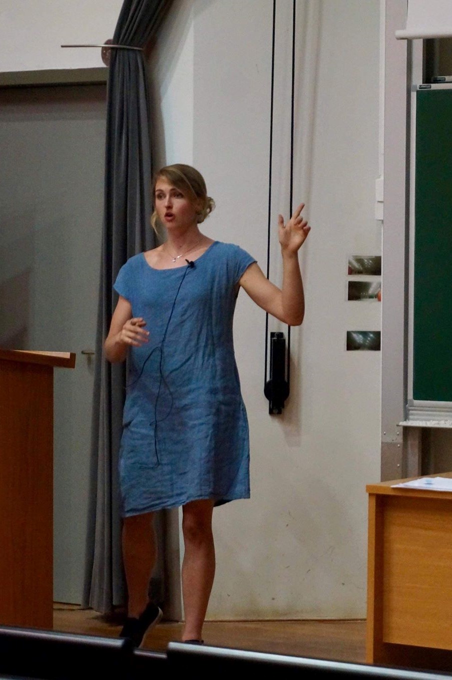
Julia Schaepe
|
Tracking Interneuron Migration in Forebrain Assembloids
Julia presented on her research building a machine learning algorithm to segment the somas of interneurons. This project is part of a pipeline that will ultimately track interneuron migration in microscopy videos, replacing and standardizing a time-consuming manual evaluation process. She implemented a UNet, a type of convolutional and deconvolutional neural network, that takes in microscopy images of interneurons and outputs a segmentation mask of the somas. Her presentation focused on developing the training and testing data sets, building the deep learning network, implementing transfer learning with a larger Kaggle cell segmentation dataset, and evaluating performance.
|
| |


